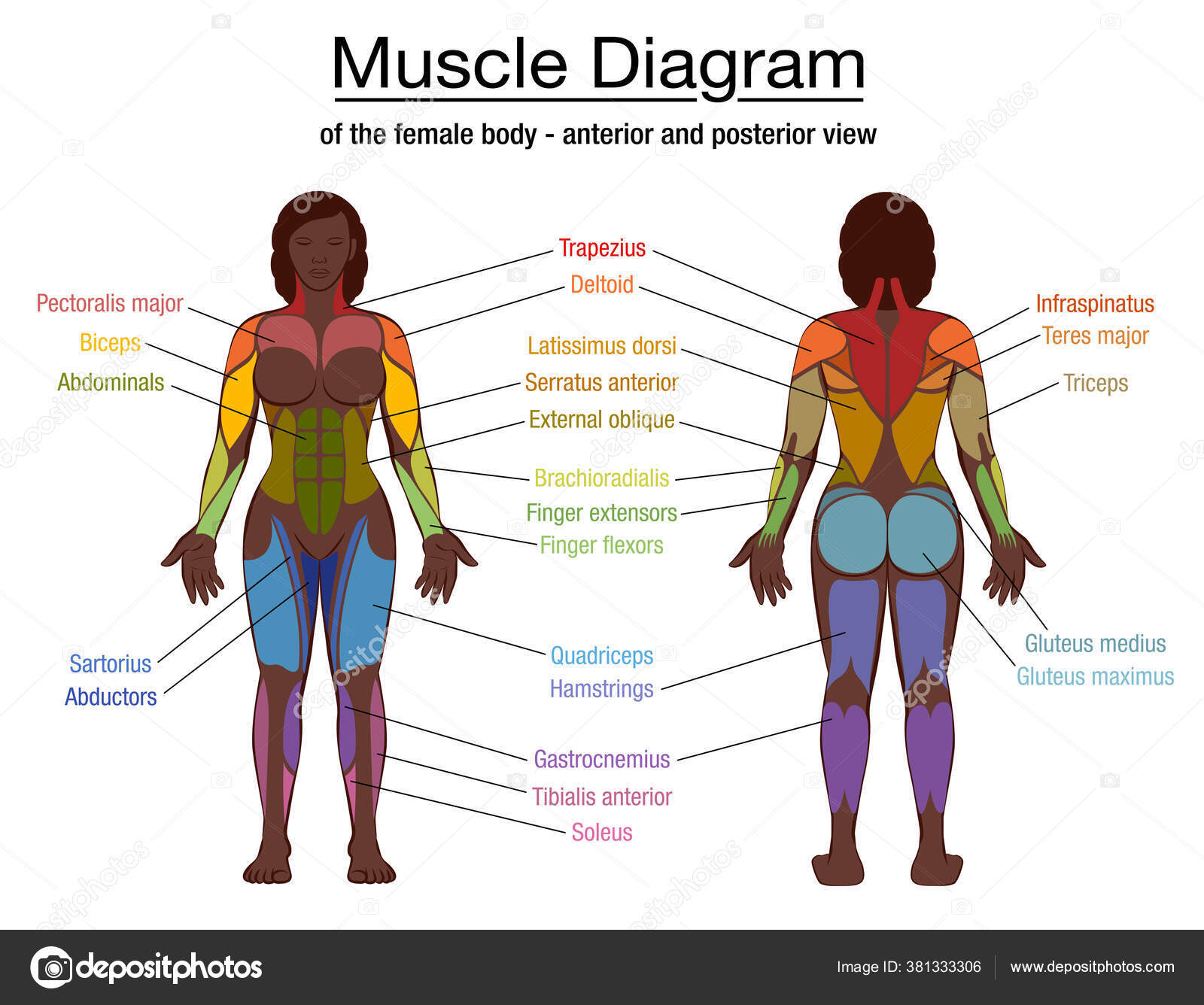
Human Muscles Diagram ~ Muscular System Anatomy Diagram Function Healthline. Visual scheme circulatory system, blood vessel. The human body consists of many muscles. This entry was posted in anatomy, muscles and tagged human muscle diagram, human muscles, muscle, muscles, muscles anatomy, muscles diagram, muscular system by admin. In this image, you will find frontalis, orbicularis oculi, zygomaticus, masseter, orbicularis oris, sternocleidomasteoid, deltoid, pectoralis major, biceps brachii, iliopsoas, adductor longus, gastrocnemius. Skeletal muscle is the only voluntary muscle tissue in the human body—it is controlled consciously.
Every physical action that a person consciously performs (e.g. Human body anatomy man and woman vector human body anatomy man and woman. This is a table of skeletal muscles of the human anatomy. The majority of muscles in the leg are considered long muscles, in that they stretch great distances. This diagram depicts human muscles.
2 555 Muscle Diagram Vectors Royalty Free Vector Muscle Diagram Images Depositphotos from st4.depositphotos.com The primary job of muscle is to move the bones of the skeleton, but muscles also enable the heart to beat and constitute the walls of other important hollow. The muscle will illuminate and the corresponding information is displayed. Almost every muscle constitutes one part of a pair of identical bilateral muscles, found on both sides, resulting in approximately 320 pairs of muscles, as presented in this article. The interactive muscle anatomy diagram shown below outlines the major superficial (i.e. Antique anatomical diagram, muscles of the human body, 19th century vintage engraving of antique anatomical diagram, muscles of the human body, 19th century human muscle stock illustrations. Muscle anatomy model 12 photos of the muscle anatomy model anatomy muscle models arm, muscle anatomy model, muscle anatomy model amazon, muscle anatomy model labeled, shoulder muscle anatomy model, human muscles, anatomy muscle models arm, muscle anatomy model, muscle anatomy model amazon, muscle anatomy model labeled. Abductors (tensor fasciae latae, gluteus medius, gluteus minimus) ilium: The trapezius or trapezoid muscles are two paired muscles that extend from the base of the thoracic vertebrae in the spine to the occipital bone and run out to the spine of the scapula.
There are three parts to the trapezius.
The primary job of muscle is to move the bones of the skeleton, but muscles also enable the heart to beat and constitute the walls of other important hollow. In this image, you will find frontalis, orbicularis oculi, zygomaticus, masseter, orbicularis oris, sternocleidomasteoid, deltoid, pectoralis major, biceps brachii, iliopsoas, adductor longus, gastrocnemius. It should be noted that there are many more muscles in the body that are not addressed by this muscle anatomy diagram, however the muscles that are of primary interest from a fitness and exercise. The muscular system is responsible for the movement of the human body. If someone wants a healthy and good life, one must understand his body. Find more reuslts at life.123.com Tapping a finger muscle, corresponding information is displayed. This diagram depicts human muscles. The trapezius or trapezoid muscles are two paired muscles that extend from the base of the thoracic vertebrae in the spine to the occipital bone and run out to the spine of the scapula. Study human anatomy 3d with reliable models & detailed articles. Broadly considered, human muscle—like the muscles of all vertebrates—is often divided into striated muscle, smooth muscle, and cardiac muscle. Almost every muscle constitutes one part of a pair of identical bilateral muscles, found on both sides, resulting in approximately 320 pairs of muscles, as presented in this article. A diagram of young and old face showing the decrease in collagen and broken elastin.
Molly smith dipcnm, mbant • reviewer: Tapping a finger muscle, corresponding information is displayed. The muscle will illuminate and the corresponding information is displayed. The human body consists of many muscles. Human muscles anatomy diagram consists of various muscles including biceps, triceps, etc.
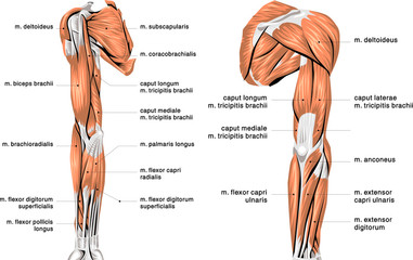
264 685 Human Muscle Wall Murals Canvas Prints Stickers Wallsheaven from t4.ftcdn.net The interactive muscle anatomy diagram shown below outlines the major superficial (i.e. Find human body muscles picture now. Muscle anatomy model 12 photos of the muscle anatomy model anatomy muscle models arm, muscle anatomy model, muscle anatomy model amazon, muscle anatomy model labeled, shoulder muscle anatomy model, human muscles, anatomy muscle models arm, muscle anatomy model, muscle anatomy model amazon, muscle anatomy model labeled. Adductors (includes madductor longus, adductor brevis, adductor magnus muscles: In this image, you will find frontalis, orbicularis oculi, zygomaticus, masseter, orbicularis oris, sternocleidomasteoid, deltoid, pectoralis major, biceps brachii, iliopsoas, adductor longus, gastrocnemius. Check out and click on the image to download it. Broadly considered, human muscle—like the muscles of all vertebrates—is often divided into striated muscle, smooth muscle, and cardiac muscle. Muscle charts of the human body for your reference value these charts show the major superficial and deep muscles of the human body.
Every physical action that a person consciously performs (e.g.
Human muscle system, the muscles of the human body that work the skeletal system, that are under voluntary control, and that are concerned with movement, posture, and balance. The human body consists of many muscles. Find the best weight lifting exercises that target each muscle or groups of muscles. Located immediately below the skin) muscles of the body. Human muscle system, the muscles of the human body that work the skeletal system, that are under voluntary control, and that are concerned with movement, posture, and balance. This application is intended to complement the study of human anatomy in medicine, biology or another discipline. A diagram of young and old face showing the decrease in collagen and broken elastin. It should be noted that there are many more muscles in the body that are not addressed by this muscle anatomy diagram, however the muscles that are of primary interest from a fitness and exercise. Body structures in full growth. This diagram depicts human muscles. The first human muscle diagram above shows you the overview of the human muscular system. Human muscles anatomy diagram consists of various muscles including biceps, triceps, etc. Speaking, walking, or writing) requires skeletal muscle.
This application is intended to complement the study of human anatomy in medicine, biology or another discipline. Superficial and deep anterior muscles of upper body superficial and deep posterior muscles of upper body. Prescription muscle relaxers have a broad ranges of uses, both for acute and chronic illness. Human muscle system, the muscles of the human body that work the skeletal system, that are under voluntary control, and that are concerned with movement, posture, and balance. Almost every muscle constitutes one part of a pair of identical bilateral muscles, found on both sides, resulting in approximately 320 pairs of muscles, as presented in this article.
Human Muscles Exercise And Muscle Royalty Free Vector Image from cdn1.vectorstock.com Muscle charts of the human body for your reference value these charts show the major superficial and deep muscles of the human body. Muscles are the only tissue in the body that has the ability to contract and therefore move the other parts of the body. The first human muscle diagram above shows you the overview of the human muscular system. Speaking, walking, or writing) requires skeletal muscle. Muscles after death, and in extreme forms of contracture in which muscle metabolism can no longer provide atp. This application is intended to complement the study of human anatomy in medicine, biology or another discipline. Find more reuslts at life.123.com Broadly considered, human muscle—like the muscles of all vertebrates—is often divided into striated muscle, smooth muscle, and cardiac muscle.
This application is intended to complement the study of human anatomy in medicine, biology or another discipline.
Muscle charts of the human body for your reference value these charts show the major superficial and deep muscles of the human body. Dimitrios mytilinaios md, phd last reviewed: Molly smith dipcnm, mbant • reviewer: The main function of the muscular system is movement. If someone wants a healthy and good life, one must understand his body. The muscular system is responsible for the movement of the human body. Check out and click on the image to download it. Broadly considered, human muscle—like the muscles of all vertebrates—is often divided into striated muscle, smooth muscle, and cardiac muscle. Learn from 3d models & articles that extend each other Connect the dots on anatomy app. Search for human body muscles picture now. You can click the links in the image, or the links below the image to find out more information on any muscle group. The muscle will illuminate and the corresponding information is displayed.